 Care
Care
Symptoms and Treatment of Canine Histiocytosis
Skin conditions in dogs are common pathological processes, and they're easy to identify since they're visible to the naked eye. An uncommon skin tumor condition in dogs is canine histiocytosis, characterized by the appearance of plaques or nodules on the skin in different areas of the body, and which can even affect internal organs.
Below, we'll describe what this process involves, how to identify it, and what treatment options are available. Ready?
What Is Canine Histiocytosis?
Histiocytosis is a pathological process in which there's excessive proliferation of certain skin cells called histiocytes.
If this uncontrolled proliferation of histiocytes occurs only in the skin, we call it cutaneous histiocytosis. If it affects other organs (liver, spleen, lungs, lymphatic system…), it's called systemic histiocytosis. In both processes, identical skin lesions develop.
The disorder in histiocyte multiplication can also lead to tumor diseases such as cutaneous histiocytoma and malignant histiocytosis (disseminated histiocytic sarcoma).
What Is a Histiocytoma?
A canine histiocytoma is a benign tumor that usually presents as a small, firm, button-shaped mass, which can ulcerate. It's usually a single, fast-growing mass that, in principle, isn't painful.
The areas most affected are the head, ears, and legs. It can resolve spontaneously within 3 months.
What Signs Does Cutaneous/Systemic Histiocytosis Cause?
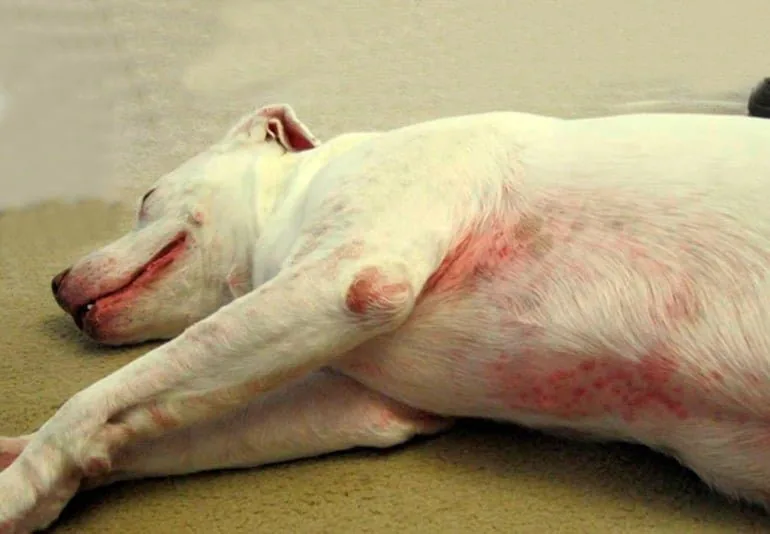
In canine histiocytosis, multiple, well-defined skin masses appear, in the form of plaques or nodules. These lesions often ulcerate and develop scabs.
With cutaneous histiocytosis, only nodules appear on the skin, most often on the head, neck, trunk, limbs, and scrotum. Systemic organs aren't affected. These lesions usually aren't painful or itchy at first, but if they ulcerate, they can become a painful process for the dog.
Systemic histiocytosis affects, besides the skin, other internal organs, so we'll see both skin symptoms and general signs: multiple skin nodules, enlarged lymph nodes, eye issues (conjunctivitis, retinal detachment, glaucoma), breathing sounds, coughing, loss of appetite, lethargy, etc.
The lesions can go through alternating periods of improvement and worsening, and can even resolve spontaneously.
Symptoms of Malignant Histiocytosis
The symptoms of malignant histiocytosis as it progresses are more serious, presenting with pale mucous membranes, weakness, difficulty breathing, and neurological signs (seizures, paresis, etc.), and masses in the liver and/or spleen. The skin and eyes usually aren't affected.
Which Dogs Are Predisposed to Histiocytosis?
- Sex predisposition: there's no sex predisposition — it can affect males and females equally.
- Age: cutaneous and systemic histiocytosis usually occurs in young or middle-aged dogs (average age 5 years). Histiocytoma especially affects dogs under 2 years old.
- Breed predisposition: systemic histiocytosis is more common in the Bernese Mountain Dog, but can affect other breeds too. The skin disease isn't limited to specific breeds. Histiocytoma is more common in the Bull Terrier, Boxer, Dachshund, Cocker Spaniel, Great Dane, and Shetland Sheepdog.
How Is Canine Histiocytosis Diagnosed?
The vet will diagnose this process through observation of clinical signs and additional tests.
To confirm that the skin lesions correspond to canine histiocytosis, a cytology and/or biopsy of the nodules/masses found on the skin or other organs needs to be performed.
In addition, an abdominal ultrasound and chest X-ray are recommended to rule out a systemic process (systemic histiocytosis affecting internal organs).
What Is the Treatment for Histiocytosis?
Currently there's no single defined treatment for this pathological process. For cases of cutaneous histiocytosis, corticosteroids are mainly used for treatment, achieving full or partial remission of skin lesions. However, recurrences are common, and ongoing treatment over time may be needed.
Other immunosuppressive medications are also used, such as cyclosporine, azathioprine, and leflunomide, which can be combined with other drugs and substances such as vitamin E and fatty acids.
In the case of histiocytoma, once the diagnosis is confirmed and a malignant tumor type has been ruled out, it's possible to wait 3 months without any therapy to see if it resolves spontaneously. If the lesion doesn't go away after this time, surgical removal or cryosurgery (burning with cold application) is recommended.
What Is the Prognosis for Canine Histiocytosis?
The prognosis for dogs with malignant histiocytosis is extremely poor, since life expectancy is usually just a few months after diagnosis.
For cutaneous and systemic histiocytosis, the prognosis depends on the response to treatment — full or partial remission of lesions can be achieved, but relapses are common and ongoing therapy may be needed.
For histiocytomas, surgical removal is generally a curative therapy that resolves the process.
Bibliography: Larry P. Tilley/ Francis W. K. Smith, Blackwell's La consulta veterinaria en 5 minutos T. Nuttall/ R. G. Harvey/ P. J. Mckeever, Enfermedades cutáneas del perro y el gato
IMPORTANT: This article is for informational purposes only and does not constitute veterinary, nutritional or behavioural advice. Every dog is different, and only your trusted vet can advise you on its specific needs.