 Care
Care
Aural Hematoma in Dogs - Is Surgery Necessary?
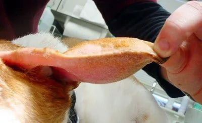
Aural hematoma is a fairly common problem in dogs, especially those with floppy ears or dogs that regularly get ear infections. It's a very easy condition to spot, since our dog's ear can end up "swelling" like a balloon.
Treatment should be as prompt as possible to prevent complications such as infections or permanent deformity of the dog's ear. Below, we'll explain the main causes, treatment, and steps to follow to prevent this ear problem in our pet.
What is an aural hematoma?
An aural hematoma is a hematoma located on the dog's ear flap — that is, a buildup of blood between the skin and the ear cartilage. A dog's ear is like a sandwich: a layer of skin, ear cartilage, and another layer of skin; and between these layers there are blood vessels.
The main problem is that there are barely any blood vessels in the dog's ear, so reabsorption of this blood can be difficult, and it ends up building up like a kind of pouch.
Initially, it's just a buildup of liquid blood and serum, but over time, if untreated, clots form that can cause permanent scarring deformities in the dog's ear and other secondary complications, such as infection of the ear cartilage (perichondritis).
How does an aural hematoma form?
Direct trauma to our dog's ear, ear scratching, or repeated head shaking can cause the blood vessels in the ear flap to rupture. This causes blood to leak out and build up under the ear's skin (between the skin and the cartilage), creating a fluid-filled swelling in the ear that varies in size depending on how much blood has leaked out.
If we let time pass without seeing the vet, new trauma to the area (such as scratching or shaking) can restart the bleeding and increase the size and severity of the condition.
What factors cause aural hematomas?
As we've seen, the cause of aural hematomas is rupture of the blood vessels in the ear flap. This rupture generally happens for 3 reasons:
- Direct trauma to the ear area (impacts, fights between dogs).
- Repeated self-trauma from scratching or head shaking due to itching or ear pain from underlying conditions (ear infections, allergies, parasites).
- Fragile blood vessels in older dogs (increasing bleeding risk even without major trauma to the area).
What factors predispose dogs to aural hematomas?
Besides those mentioned, these factors can make your dog more prone to developing an aural hematoma.
- Ear type: aural hematomas are more common in dogs with floppy ears.
- Sex: aural hematomas occur more often in males than females.
- Age: the incidence of aural hematomas increases with age, due to fragile blood vessels in older dogs.
- Recurrence: aural hematomas can leave lasting effects in the area, and recurrence is common in dogs who've had this condition before.
- Underlying conditions: dogs with conditions causing pain or itching in the ear area are more prone to developing aural hematomas from repeated self-trauma (scratching or shaking).
How is an aural hematoma diagnosed?
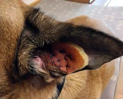
Seeing a fluid-filled swelling on a dog's ear flap is a clear sign of an aural hematoma. Diagnosis is confirmed by draining/emptying its contents via needle aspiration, confirming it's blood. This should always be done by a vet.
What is the treatment for an aural hematoma?
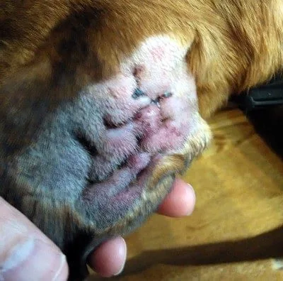
For treatment of an aural hematoma to be successful and not recur, the first thing the vet needs to determine is the underlying cause behind it and treat it (ear infection, allergy, parasites); since if trauma to the ear area continues, even after removing the hematoma, it will come back.
Not every aural hematoma is caused by an underlying ear infection or itching. There can be capillary fragility or trauma as the main cause, and in these cases, the step to take after removing the hematoma is to take extra precautions to prevent future trauma to the ears.
To remove the aural hematoma itself, the following veterinary procedure needs to be followed:
- If it's a small aural hematoma and not a recurrence, draining its contents via needle aspiration may resolve the problem. Corticosteroids and antibiotics may be needed to prevent complications.
- If it's a more severe aural hematoma or a recurrence, surgery is needed to resolve it. The technique involves making an incision to fully drain the hematoma and thoroughly clean the ear flap and external ear canal. The incision made is a special type (the edges are trimmed) to keep it from closing in the following days, allowing the wound to keep draining. Over the area that was affected by the hematoma, stitches are placed parallel to the ear's long axis (several stitches are given), eliminating the dead space where the fluid had collected and keeping the affected skin in contact with the ear cartilage. Stitches can be placed directly on the skin with no additional support, or surgical sponges can be used, which also absorb any secretions. The wound should always be protected, at least for the first 48 hours, with a bandage. Corticosteroids and antibiotics may be needed.
What steps should be followed after aural hematoma surgery?
Flawless surgery can turn into a failure if we don't strictly follow the post-op care instructions.
Besides following the medication schedule and incision care set by the vet, the main thing is to keep the dog from scratching or bumping the wound, to prevent renewed bleeding and allow the ear to heal properly. To do this, our dog needs to wear an Elizabethan collar until the stitches are removed (10-15 days after surgery).
Anti-inflammatory medication (corticosteroids) helps control any pain or itching from the procedure, so the dog will be comfortable on that front; so we'll mainly need to keep them from playing roughly or bumping into furniture around the house if they're not used to moving around with the Elizabethan collar.
IMPORTANT: This article is for informational purposes only and does not constitute veterinary, nutritional or behavioural advice. Every dog is different, and only your trusted vet can advise you on its specific needs.