 Care
Care
Conjunctivitis in Dogs: Symptoms, Diagnosis, and Treatment
Conjunctivitis is inflammation of the conjunctiva, a transparent mucous membrane that lines the inner part of the eyelids and the white surface, or sclera, of the eyeball. It can have a primary or secondary cause.
It's a fairly common reason for a vet visit and can stem from different causes. It requires veterinary attention and proper eye treatment to clear up. Let's take a closer look at everything related to this condition.
Causes of Conjunctivitis
We've already described what conjunctivitis is. In this section, we'll focus on its causes or etiology. Usually only one of these causes is present, though it's possible, if less likely, for several to occur together. These are:
- Allergic conjunctivitis: usually associated with atopy and food allergies, so it's not uncommon to find systemic signs as well. However, conjunctivitis can also be the only manifestation of an allergic process.
- Plasmacytic conjunctivitis: this type of conjunctivitis involves an infiltration (buildup) of lymphocytes and plasma cells in the conjunctiva. This spreads as a grayish membrane over the edge of the third eyelid.
- Bacterial conjunctivitis: usually secondary to abnormalities or malformations (entropion, ectropion) of the eyelids or eyelashes, trauma, or chronic skin conditions.
- Systemic diseases: many systemic diseases present with conjunctivitis, such as canine distemper, ehrlichiosis, or leishmaniasis.
Symptoms of Conjunctivitis in Dogs
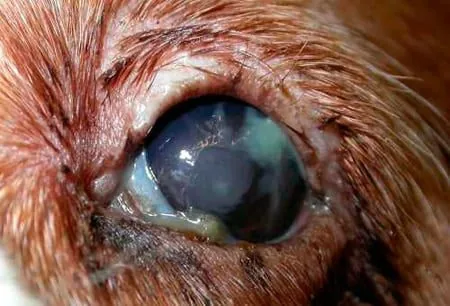
Conjunctivitis can appear in one or both eyes, depending on the cause. It's generally an uncomfortable condition, so we may notice the dog keeping the eye closed, although as mentioned, it can be linked to other conditions with several symptoms showing up at once. The symptoms of conjunctivitis include:
- Conjunctival hyperemia, or redness of the eye.
- Discharge or exudate, which can be clear and watery, mucous, or purulent.
- Chemosis, or a swollen, fluid-filled (edematous) conjunctiva.
- Loose folds forming in the conjunctival fornices.
- Other symptoms may appear depending on the type of conjunctivitis the dog has.
Diagnosing Conjunctivitis
The vet will determine whether the hyperemia (red eye) is due to inflammation of the conjunctiva (conjunctivitis) or episcleral inflammation (episcleral vascularization). By instilling a drop of phenylephrine, this can be determined, since in cases of conjunctivitis, the hyperemia will largely disappear as the tissue turns pale. Otherwise, the inflammation or hyperemia will persist. This could point to glaucoma or uveitis.
A differential diagnosis must be made against five conditions that can mimic conjunctivitis. These are: keratitis (ulcerative or non-ulcerative), dry eye (keratoconjunctivitis sicca), scleritis, uveitis, and glaucoma.
To rule these out, the vet can run additional tests or exams at the clinic. These include:
- Fluorescein test, which stains the stroma or the depth reached by an ulcer, telling us whether one is present.
- Tonometry: measures intraocular pressure with an instrument. This should read between 15 mmHg and 25 mmHg. Results below 10 point to uveitis, and above 30 to glaucoma. A difference of more than 5 mmHg between the two eyes should be considered significant.
- Schirmer test: quantitatively evaluates tear production in each eye. Special strips of paper, folded at one end, are placed in the lower conjunctival sac while the animal keeps its eyes closed. After one minute, the result is read. Normal results are 15-20 mm of moisture in a minute. Values below 10 mm suggest keratoconjunctivitis sicca. Below 5 mm indicates the same, but more severe.
Treatment for Conjunctivitis
The most important thing is to find the underlying cause so it can be corrected, since treatment depends on that etiology. Only the vet can tell us which type of conjunctivitis we're dealing with. Any eyelid abnormalities need to be corrected, any systemic disease treated, and a topical treatment applied to resolve the conjunctival inflammation.
Whenever a primary or secondary bacterial cause is suspected, broad-spectrum topical antibiotics should be used, for example a poly-antibiotic eye drop containing neomycin, polymyxin B, and bacitracin. The frequency of application depends on how severe the conjunctivitis is.
If needed, a culture and antibiogram should be performed. In cases of chlamydial conjunctivitis, topical chloramphenicol or tetracycline should be given. For follicular conjunctivitis, as with plasmacytic conjunctivitis, a topical corticosteroid (0.1% dexamethasone) should be used.
Combining topical cyclosporine (0.5-2%) with topical corticosteroids is very beneficial for a faster resolution of symptoms.
Eye Hygiene for Conjunctivitis
To apply any medication properly, the eye needs to be as clean as possible. This ensures the medication penetrates well and works effectively. So, before administering medication, all discharge and crust needs to be removed. With the eye as clean as possible, it's time to apply the medication prescribed by the vet. Repeat the cleaning before each application as long as discharge is present.
We can do this by moistening a piece of gauze or cotton with saline solution or, simply, water. Clean gently and superficially from inside out, using a different piece of gauze for each eye. Remember that conjunctivitis in one eye can spread to the healthy one.
It doesn't hurt to use over-the-counter artificial tears or lubricating ointments between doses of the prescribed eye drops, to keep the eye moisturized and lubricated. It's important not to give your dog any eye drops of your own choosing at home. This could worsen the conjunctivitis and lead to further symptoms. Always consult your vet to ensure your animal's wellbeing.
IMPORTANT: This article is for informational purposes only and does not constitute veterinary, nutritional or behavioural advice. Every dog is different, and only your trusted vet can advise you on its specific needs.