 Care
Care
The Third Eyelid in Dogs - Issues and Treatments
Unlike people, dogs have three eyelids — two of them are covered in fur and visible to the naked eye (upper and lower), while the third is "hidden" beneath the eyelids and isn't visible under normal conditions.
Sometimes, this third eyelid can become visible, appearing from the tear-duct area as a thin, pinkish-white membrane covering part of the eye's surface. Let's look at the main problems this third eyelid can cause and how they're treated.
What is a dog's third eyelid?
Besides the two visible eyelids (upper and lower), dogs have a third eyelid called the nictitating membrane.
This extra eyelid is a thin membrane of connective tissue located at the inner corner of the eye, hidden beneath the eyelids. It's not covered in fur like the other two eyelids, nor does it have muscle, but it does contain a tear gland called the gland of the third eyelid (Harder's gland).
Under normal conditions, a dog's third eyelid isn't visible, but it can show under certain circumstances, appearing as a pinkish-white membrane covering part of the eye's surface.
What is a dog's third eyelid for?
The third eyelid, or nictitating membrane, has two main functions in dogs:
- Protective function: When a foreign particle enters a dog's eye, the eyeball retracts into the socket and the third eyelid appears, covering part of its surface; this membrane can help remove foreign objects through a sweeping effect, like a windshield wiper, acting as a reflex to prevent eye damage. It can also show if there's an eye issue, such as conjunctivitis or ulcers, acting as a protective layer until the issue resolves.
- Hydrating function: The third eyelid contains a tear gland (the gland of the third eyelid) responsible for around 40% of total tear production, making it essential for keeping the eye properly hydrated.

When does the third eyelid become visible?
As we've seen, the nictitating membrane shows up, covering the eye's surface, as a defense mechanism against irritation or issues that could damage the eyes.
So if we notice the third eyelid in our dog, it's very likely they have a foreign particle in their eye, or an existing eye issue/injury already present (conjunctivitis, ulcer, etc.).
It's also common to see the third eyelid show up in dogs that feel unwell or have some general condition (an infection, gastroenteritis, fever, etc.).
That's why the third eyelid can serve as an "alarm" pointing to our dog possibly having an eye issue or a general health problem.
What are the main third eyelid issues in dogs?
We can mainly describe three conditions affecting the nictitating membrane in dogs:
- Third eyelid protrusion.
- Prolapse of the third eyelid gland, or cherry eye.
- Third eyelid tumors.
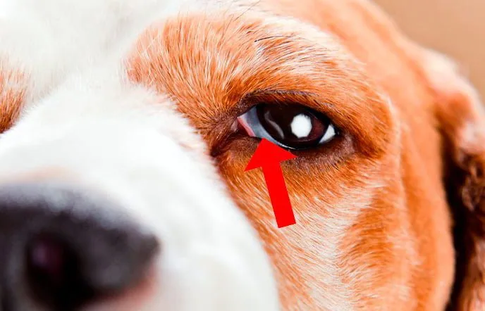 Third eyelid protrusion
What does third eyelid protrusion involve?
Under normal conditions, the nictitating membrane isn't visible, and it shows up as a defense mechanism in response to eye injuries or general health conditions such as fevers, gastrointestinal issues, etc.
If we notice a pinkish-white membrane appearing in our dog's eye(s) from the inner corner (near the tear duct), covering part of the eye's surface, and up to 6 hours pass without that membrane going back to its place and stopping being visible, we should check with the vet. Although it's not an emergency, it can point to an eye issue or even an underlying general illness in our pet.
What does prolapse of the third eyelid gland involve?
At its base, the nictitating membrane contains a tear-secreting gland (the gland of the third eyelid), held in place by bands of connective tissue.
If this attachment is weak, the gland can prolapse outward, appearing as a small pink lump (mass) near the tear-duct area of the eye.
This type of issue is known as cherry eye, and it's a fairly common condition seen at the vet clinic. Although it can occur in any dog, certain breeds are predisposed to this issue: Bulldog, Boxer, Bloodhound, Beagle, Pekingese, Chihuahua, Cocker Spaniel, among others.
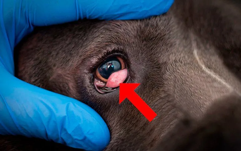 Prolapse of the third eyelid gland
This eye issue needs to be treated by a vet as soon as possible, since it can cause eye problems such as dry eye (keratoconjunctivitis sicca), since tear production is affected.
Initially, a topical anti-inflammatory treatment is used (corticosteroid eye drops) to reduce swelling in the affected area. Afterward, under anesthesia, the prolapse is reduced (the gland is put back in place) and the gland is stitched in place to keep it from prolapsing again.
Once the procedure is done, antibiotic and anti-inflammatory therapy needs to be given through eye drops, and the animal should avoid straining in ways that could put tension on the stitched area. It's essential to use an Elizabethan collar to keep the dog from scratching the area until they've fully recovered.
Third eyelid tumors
Although tumors of the nictitating membrane aren't very common, they can occur, especially in large, older dogs. The most common tumor is melanoma, which tends to be malignant with a fairly high likelihood of metastasis. Tumors can also affect the third eyelid gland, in which case they're called adenocarcinomas.
Tumors are diagnosed via fine needle aspiration to analyze the cells involved, and treatment involves complete surgical removal of the nictitating membrane. Supportive techniques such as cryotherapy, laser, or radiotherapy can also be used.
As we can see, the third eyelid plays a very important role in our pet's eye health, so if we notice any of these issues in our dog, we shouldn't let them slide — we should see the vet as soon as possible, since early treatment is key to preventing serious eye damage.
Bibliography Carlson and Giffin. 2002. Manual práctico de veterinaria canina. Madrid. Editorial el Drac.
IMPORTANT: This article is for informational purposes only and does not constitute veterinary, nutritional or behavioural advice. Every dog is different, and only your trusted vet can advise you on its specific needs.