 Care
Care
Why Does My Dog Have White Eyes?
On the outside, dogs' eyes are fairly similar to ours. Most are brown, in every shade, though some dogs have blue or amber eyes, and some even have one eye of each color.
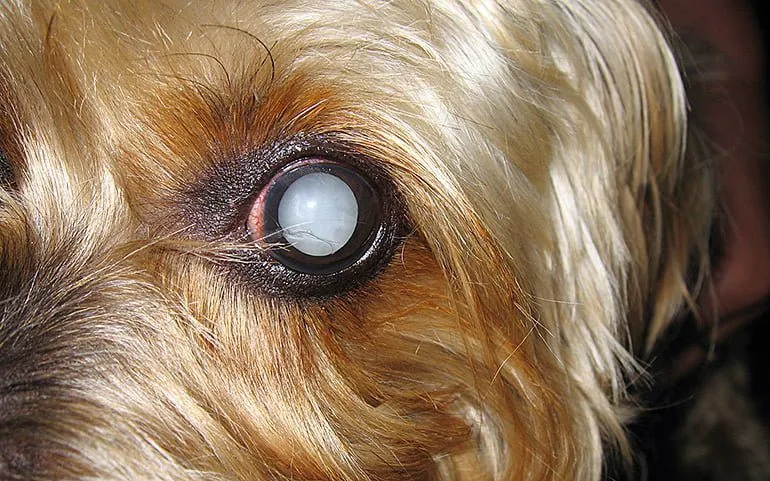
But sometimes, our dog's natural eye color changes and turns whitish. We may notice a white spot, or a kind of film or layer clouding the whole eye. Below, we'll explain what causes white eyes in a dog.
Most common symptoms of eye conditions
First, it's worth noting that sometimes, when dogs are falling asleep or even while sleeping, their eyes can roll back and show white. In this case, it's not due to any condition.
It's simply the closed eye visible when the eyelids part slightly, for some reason, usually posture or an attempt to stay awake. Of course, it isn't a health concern at all.
On the other hand, noticing a whitish tone on the open eye is a sign of an eye condition. There are different situations here, since a spot isn't the same as an opaque film covering the eye fully or partially:
- Film over the eye: a whitish, opaque membrane extending from inside the eye is known as the nictitating membrane, or third eyelid.
- Cloudiness in the eye: cloudiness refers to the eye losing its transparency. It can come with other symptoms, such as pain.
What is the third eyelid in dogs?
This is a structure that can prolapse and cover the eye fully or partially, giving the impression that the dog's eyes are rolled back, as if the eye had turned inside the socket. It's a third eyelid with very important functions. It's located at the inner corner of the eye, is opaque, and normally not visible.
It contains a tear gland responsible for producing almost half of the total volume of tears. There are several causes that can make this membrane show, such as eye pain, retraction of the eye, cartilage eversion, or a foreign object. We should contact the vet.
One notable condition is known as third eyelid paresis. This involves the eyelid protruding over the eye. In some dogs, it's a birth defect. It's not a clinical problem — it would only matter cosmetically for a dog being shown in beauty competitions. No treatment is needed.
Corneal injuries in dogs
The cornea is the clear part of the eye. It's covered by a surface layer of epithelial cells, with a middle and inner layer as well. Any injury causes a wound that inflames the damaged area.
We sometimes notice this inflammation as a blurry, opaque, whitish area on the eye. If we don't see the injury but notice a lot of pain, crossed eyes, tearing, light sensitivity, and the third eyelid prolapsed to protect the damaged eye, we should see the vet. Prompt attention is needed.
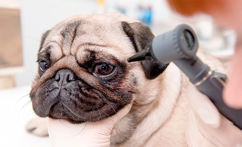
Corneal injuries are more common in dogs with prominent eyes, such as the Pekingese or Pug, though any dog can suffer them, regardless of breed. The main ones are:
- Corneal abrasion: even just an eyelash growing toward the cornea can cause an abrasion. The same happens with a foreign object lodged in the eye. A vet exam and medication are needed to prevent infection and reduce pain.
- Corneal ulcer: similar to an abrasion but deeper. It usually results from trauma, though it's sometimes linked to other eye conditions or diseases such as diabetes, hypothyroidism, or Addison's disease. Without treatment, the eye can be lost. Surgery is sometimes needed.
- Corneal dystrophies: hereditary diseases of the cornea in both eyes, appearing as an oval or rounded grayish-white opacity. They can cause blindness. They can be surgically removed, but will form again.
Types of canine keratitis
Noticing cloudiness over the eye can point to keratitis. This is inflammation of the cornea that makes it cloudy. There are also symptoms such as intense tearing, crossed eyes, light sensitivity, or the third eyelid protruding. The dog will rub at their eye with their paws.
There are several types of keratitis. What they all have in common is that, if untreated, they can end up causing partial or total blindness. So, a vet visit is essential. Here are the main types of keratitis:
- Ulcerative keratitis: occurs as a complication of a corneal ulcer or another condition called dry eye (keratoconjunctivitis sicca). The cornea takes on a milky white color.
- Infectious keratitis: here, the complication is due to the presence of bacteria, triggering a pus-filled eye discharge. The eyelids can become swollen. In a smaller percentage of cases, the infection is caused by fungi.
- Interstitial keratitis: here, the cloudiness seen over the eye has a bluish-white tone. It's caused by the infectious hepatitis virus. It may clear up within a few weeks, or remain as a permanent aftereffect of this disease.
- Vascular and pigmentary keratitis: loss of transparency here is due to blood vessels and connective tissue growing into the eye. In the pigmentary form, melanin is deposited on the cornea. One specific type is pannus. It can't be cured, but treatment can stop it from progressing.
What are cataracts?
Cataracts show up in the eye as a milky gray film. Although they're usually linked to aging, the truth is there are also juvenile cataracts of congenital origin. These appear before six years of age and in both eyes, though not always at the same time. Acquired cataracts, on the other hand, are the ones affecting older dogs.
These are also the ones that appear as a complication of diabetes. This type of cataract starts in the center of the eye and spreads outward. A cataract is considered mature once the whole lens appears opaque. Depending on each case, the vet will assess whether surgery is needed.
What is nuclear sclerosis in dogs?
This is a natural degeneration of the lens that occurs with aging. It's not a disease. It happens because new fibers keep forming continuously around the outer edge of the lens, pressing toward its center. The result is a bluish haze that won't interfere with vision. Its appearance can make it easy to confuse with cataracts.
Glaucoma in dogs
Glaucoma is a serious condition that can lead to blindness. It occurs when more aqueous humor forms than is drained, raising pressure inside the eye. This increase can damage both the optic nerve and the retina.
Depending on its origin, a distinction is made between primary glaucoma, which is hereditary, and secondary glaucoma, resulting from another disease. It causes pain, tearing, crossed eyes, hardness to the touch, a fixed, whitish gaze, and a larger pupil. It's a veterinary emergency — within hours the dog could go blind. Drug treatment exists, and surgery may also be an option.
Bibliography Carlson and Giffin. 2002. Manual práctico de veterinaria canina. Madrid. Editorial el Drac.
IMPORTANT: This article is for informational purposes only and does not constitute veterinary, nutritional or behavioural advice. Every dog is different, and only your trusted vet can advise you on its specific needs.